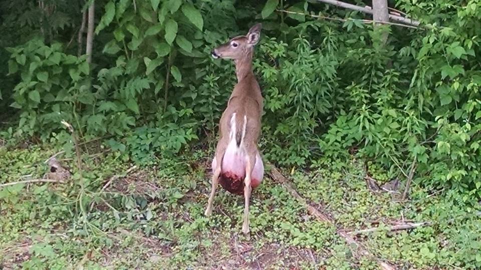
In the case of the pot-bellied deer, there are several possibilities of what might be going on (we call these “rule-outs”).
Here are the rule-outs that top the list:
- Hernia. A hernia occurs when internal organs or tissues pop through an opening in the muscular wall that surrounds the abdomen but are still covered in skin. This can happen in young animals if there are “holes” in the abdomen at the time of birth. Similarly, in adult animals hernias occur when there is a tear in the muscular wall. The tear most often is a result of trauma (fighting or being hit by a car) or straining and pressure associated with parturition (giving birth).
- Mastitis. Mastitis is a inflammation of the mammary tissues or udders. This is most often associated with microscopic bacteria in the environment or on the skin of the deer that invade mammary tissue.
- Abscess. Abscesses are collections of pus that get walled off as an inflammatory reaction in the deer’s body. Abscesses are associated with bacteria that invade the body of the deer and they can form in just about any tissue or organ. We can see them on the surface of the deer when bacteria get past the natural protective barriers of the skin; most often through cuts or other breaks in the skin. When cut into, abscesses can range from the consistency of pudding to cottage cheese, are tan, yellow, or green, and often have a foul odor.
- Hematoma. A hematoma is simply a fancy term for a collection of clotted blood. We can see them with any sort of trauma or disease that could result in bleeding.
- Tumor. There are a variety of tumors that can lead to masses on the surface deer. The most common tumor that occurs on the body of deer is a fibroma, which is associated with a viral infection. However, these typically result in variably-sized, firm, black, masses on the skin, which is quite different from what we see in this deer. A variety of other tumors can occur on the body of deer but are rare, including bone tumors, antler tumors, and other skin tumors.
Based on the enormous size of this tumor, its location, and the fact that the deer is not showing other outward signs of disease, the most likely cause of this mass is a hernia.
Hernias may not result in clinical signs other than the enlargement unless the protruding organs twist or are pinched and damaged. Based on the time of year and the fact that it is a doe, potential causes of the hernia include trauma (being hit by a car) or parturition. The bottom surface of the mass is covered with blood, which likely resulted from the mass being dragged on the ground, and that could be seen with any of the rule-outs listed above.
If this is a hernia, the long-term prognosis for this deer is not good as the protruding organs and tissues are unlikely to go back to their normal position in the abdomen and the muscular wall to heal. While the hernia will only result in severe disease if the internal organs are pinched or damaged or if secondary infections occur, the location and size of the mass will indirectly impact her ability to move.
None of the listed rule-outs have population impacts for wild white-tailed deer.
As with all of our photo diagnoses, the only way to confirm them would be to physically exam the deer. Most of these potential diagnoses could easily be confirmed or ruled-out by cutting into the abdominal mass and examining the contents (i.e. is there blood, tissue, pus, or internal organs).
-Dr. Justin Brown, PGC Vet
(and his lovely assistant J.T. Fleegle)
If you would like to receive email alerts of new blog posts, subscribe here.
And Follow us on Twitter @WTDresearch
